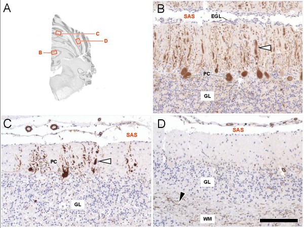
Figure 3.
Spatial characteristics of cerebellar cortical degeneration in the β-III spectrin deficient beagle after calbindin-immunohistochemistry and haematoxylin counterstain.(A) Navigator figure depicting the sampled areas. (B) The ventral aspects of the vermis show least numeric loss of calbindin-positive (brown) Purkinje cells (PC), a narrow subarachnoid space (SAS) and remnants of the external germinative cell layer (EGL). Furthermore, the granule layer (GL) is well populated and its histoarchitecture is preserved. Degenerative changes are restricted to dystrophic dendrites (white arrowhead). Purkinje cell loss becomes increasingly evident in the dorsal vermis (C) and the ansiforme lobulus of the cerebellar hemispheres (D). Resident PC show thickening and abnormal arborisation (C, white arrowhead) of the dendrites. Concomitantly the EGL is cytodepleted and the granular layer becomes mildly disorganised. Calbindin-staining in the most affected hemispheres is restricted to scattered axons, loosely bundled in the foliary white matter (D, black arrowhead). PC perikarya in many folia are completely missing while the Purkinje cell layer features a moderate Bergmann´s gliosis. Scale bar: 0.7 cm for A; 130 μm for B,C,D.

