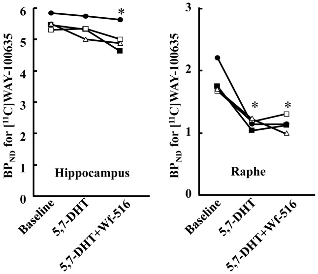
Figure 7. Alteration of BPND for [11C]WAY-100635 in rats treated with a toxicant for 5-HT neurons, 5,7-DHT.
Four rats each underwent three [11C]WAY-100635-PET scans, at baseline, after 5,7-DHT treatment, and after oral administration of 30 mg/kg Wf-516 (5,7-DHT + Wf-516), in the indicated chronological order. Each symbol represents individual BPND in the hippocampus (left) and raphe nucleus (right). These changes of BPND in each region were statistically examined by one-way repeated-measures ANOVA followed by least significant difference test. *p<0.05 compared with each baseline.

