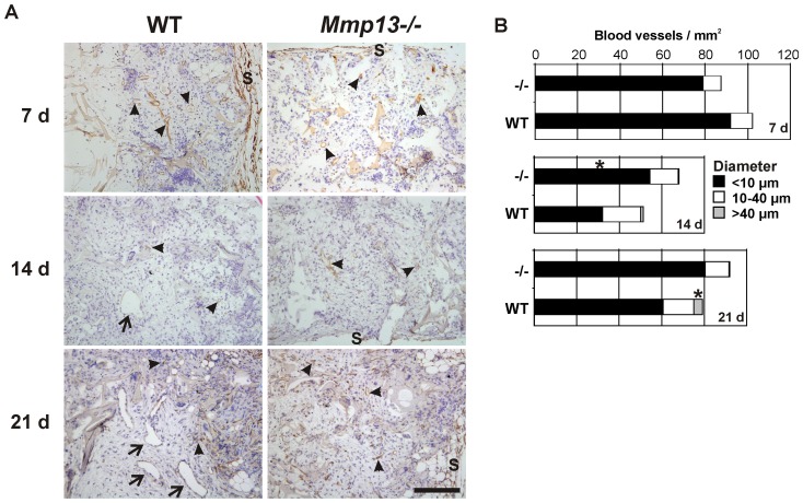
Figure 3. Altered vascular pattern in granulation tissue of Mmp13−/− mice.
(A) Sections of experimental granulation tissue of wild-type (WT) and MMP-13 knockout (Mmp13−/−) mice harvested at indicated time points were immunostained for blood vessels using CD34 as a marker. The arrowheads indicate microvessels and medium sized vessels (diameter<40 µm) and arrows indicate large vessel structures (diameter>40 µm). (s, implant surface; scale bar = 200 µm. (B) The number and the diameter of CD34-positive blood vessels were determined in defined areas of cellular granulation tissues with digital image analysis. *Statistically significant difference in the density of microvessels (<10 µm) at 14 d and of the large vessels (>40 µm) at 21 d (P<0.05, MannWhitney U test, n = 5–6).

