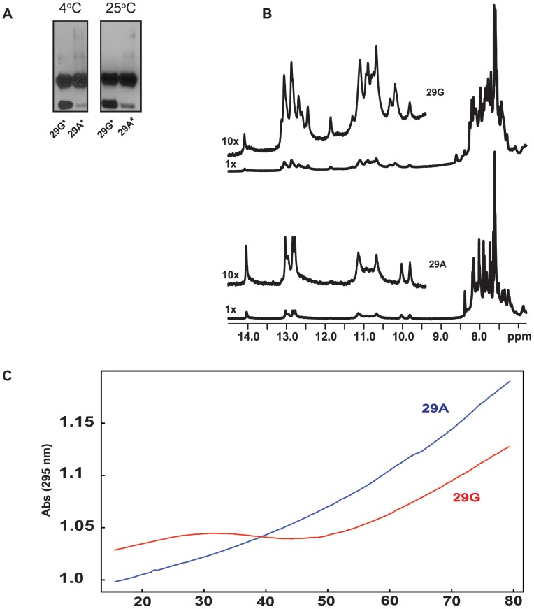
Figure 4. Higher-Order Structure in Duplex and Single Stranded Oligonucleotides.
(A) 15% non-denaturing polyacrylamide gel electrophoresis of duplex oligos in the presence of 40 mM NaCl. The oligos were incubated in binding buffer (Materials and Methods) for 40 min and 20 min at 4°C and 25°C respectively. 29G* (Lanes1 and 3), 29A* (Lanes 2 and 4) are at 4°C and 25°C respectively. (B) 1D proton NMR spectra (imino, amino and aromatic signal regions) of oligonucleotides 29G (top) and 29A (bottom) in the presence of 40 mM Na+, 10 mM phosphate, pH 7.0. (C) Denaturation profiles of single stranded oligos 29G (red) and 29A (blue) at 295 nm wavelength. Experimental conditions: 40 mM Na+, 10 mM phosphate, pH 7.0.

