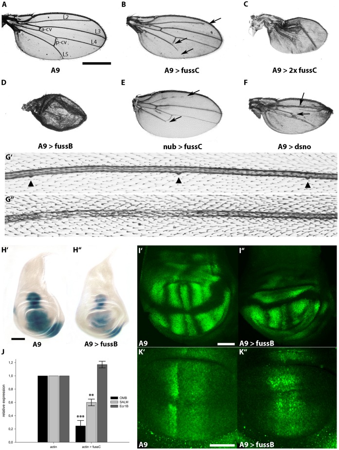
Figure 3. Ectopic expression of fussel in the wing disc reduces wing size, leads to loss of veins, loss of campaniform sensilla and interferes with the expression of BMP target genes.
(A) Control wing of a male fly from the A9-Gal line. Longitudinal (L2 to L5) and cross veins (a-cv, p-cv) are indicated. (B) A9-Gal4; UAS-fussC. The wing is smaller, arrows indicate the truncation of L2, L5 and the p-cv. (C) A9-Gal4; UAS-fussC/UAS-fussC. Expression of two copies of UAS-fussC enhances the observed phenotype. (D) A9-Gal4; UAS-fussB. Expression of one copy of fussB leads to a reduction of wing size and a severe disruption of the overall wing structure. (E) nub-Gal4; UAS-fussC. The L2 and L5 veins are truncated. (F) A9-Gal4; UAS-dSno. Expression of dSno leads to a reduction of wing size and a loss of the L4 vein. (G) Mis-expression of fuss leads to loss of campaniform sensilla. (G’) Medial part of the L3 vein of a male A9-Gal4 fly. Three campaniform sensilla are marked with arrowheads (G”) A9-Gal4; UAS-fussC. Distal to the p-cv, all campaniform sensilla are lost. (H) fuss represses omb expression. Micrographs of X- Gal- stained female L3- wing discs: (H’) omb-lacZ; UAS-fussB; (H”) omb-lacZ/A9-Gal4; UAS-fussB/+. (I) The blistered (dSRF) domain in male L3-wing discs is disrupted by fuss expression. Confocal scans of (I’) A9-Gal4; (I”) A9-Gal4; UAS-fussB. (J) Relative expression of omb, salm and ecr1b in actin-Gal4/fussC L3-larvae compared to actin-Gal4 controls. Asterisks indicate the level of statistical significance (t-test **p<0.01, ***p<0.001). (K) fuss disrupts the pattern of activated Mad but does not inhibit its phosphorylation. Confocal scans of anti-phospho-SMAD1/5 stained L3-wing discs: (K’) A9-Gal4; (K”) A9-Gal4; UAS-fussB. Scale bars represent 500 µm (A), 100 µm(H’) and 50 µm(I’, K’).

