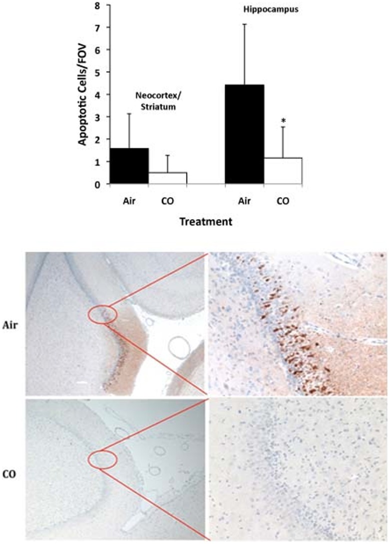
Figure 6. (TUNEL). Quantitation of TUNEL staining in indicated brain sections from neonatal piglet after CPB ± CO showed more apoptosis in the control group versus CO in the hippocampus and trending sections from the neocortex and striatum.
Data represent mean ± SD of 6 animals/group, *p<0.03. Untreated pigs showed <0.1 positively stained cell/field of view (FOV). (Caspase-3). Representative tissue sections stained for activated Caspase-3 in the hippocampus from neonatal piglets after CPB ± CO. Note the intense positive brown staining indicating activated caspase-3 localized to the hippocampus in the control, air-treated pigs versus nearly no positive staining in the CO treated pigs. Images are representative of n = 6–8/group.

