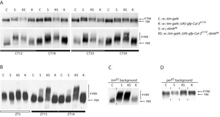
Figure 6. PER and TIM proteins in flies Cul-3K717R and slmbm flies.
(A) Anti-TIM (top) and anti-PER (bottom) Western blots of head extracts. Flies were entrained 3 d in LD and transferred to DD for collection during the first and second day of DD. Gray and black bars indicate subjective day and subjective night, respectively. (B) Anti-PER Western blots of head extracts. Flies were entrained 3 d in LD and collected the fourth day. White and black bars indicate day and night, respectively. (C, D) Anti-PER (C) and anti-TIM (D) Western blots of head extracts tim0 and per0 backgrounds, respectively. Flies were entrained for 3 d in LD and transferred to DD for collection at CT27. Vertical dashes indicate that the two surrounding slots were not contiguous on the gel. Two to three independent Western blots were done for each condition with very similar results.

