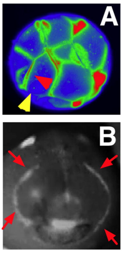
Figure 2. Voltage gradients in vivo.

A: Fluorescent voltage reporter dyes allow characterization of physiological gradients in vivo, such as this image of a 16-cell frog embryo that simultaneously reveals cells’ transmembrane potential levels (blue = hyperpolarized, red = depolarized) in vivo, as well as domains of distinct Vmem around a single blastomere’s surface (compare the side indicated by the yellow arrowhead with the one indicated by the red arrowhead). Provided courtesy of Dany Adams.
B: Isopotential cell fields can also demarcate subtle prepatterns existing in tissues, such as the hyperpolarized domains (red arrowheads) that presage the expression of regulatory genes such as Frizzled during frog embryo craniofacial development; these patterns of transmembrane potential are not merely readouts of cell state but are functional determinants of gene expression and anatomy (Vandenberg et al., 2011).
