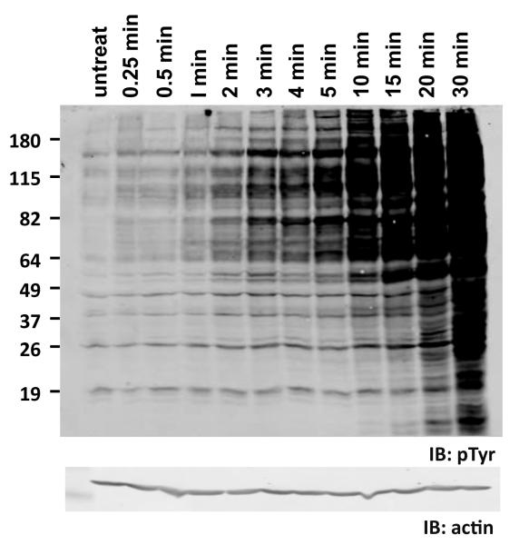
Figure 2. Constitutive rate of tyrosine dephosphorylation in cells is high.
Human 293T cells were treated with 50 micromolar pervanadate for the indicated times to inhibit cellular tyrosine phosphatases. Cell lysates were then immunoblotted with anti-phosphotyrosine antibody (pTyr). Levels of tyrosine phosphorylation rise rapidly in pervanadate-treated cells. Lysates were also immunoblotted with anti-actin as a loading control (bottom). Positions of molecular weight markers are indicated on left (in kDa).

