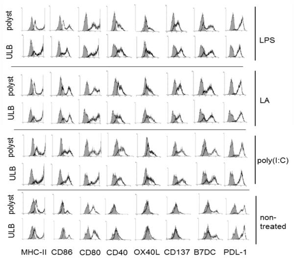
Figure 1.
Expression of costimulatory molecules by mature mDCs. Flow cytometry analysis of costimulatory molecules on mDCs cultured for 48 h on ULB or polystyrene surfaces in the presence of lipoteichoic acid (TLR-2 ligand), poly(I:C) (TLR-3 ligand), ultrapure LPS (TLR-4 ligand) or left untreated. Grey histograms represent isotype controls. An experiment representative of 4 independent experiments is depicted.

