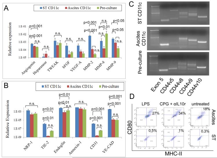
Figure 6.
Angiogenic and immunological properties of solid tumor and ascites CD11c cells. Solid tumor and ascites CD11c cells were isolated by immunomagnetic purification, and RNA was extracted and reverse-transcribed. Then, quantitative real-time PCR was performed to analyze the expression of several angiogenic molecules (A) and endothelial markers (B) in these cells. Data were analyzed by ANOVA followed by followed by Tukey-Kramer Multiple Comparisons post-Test. An experiment representative of 2 independent experiments is shown. (C) Expression of different CD44 variants by the same cells was analyzed by qualitative PCR analysis. (D) CD11c cells isolated as above from solid tumor and ovarian cancer ascites were cultured for 1 week in RPMI 10% FBS containing LPS; CPG plus anti-IL10 receptor; or left untreated. Then expression of CD80 and MHC-II was analyzed in these cells by flow cytometry. Live CD11c cells isolated from 4 independent experiments were pooled and run in quadruplicate for this study.

