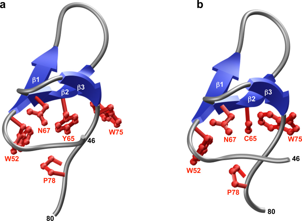
Figure 3.
3D atomic models of wild type (A) and Y65C-mutant (B) WW domains of human PQBP1. The triple-stranded β-sheet of the WW domains is shown in blue and the intervening loops in gray. The sidechains of residues W52, Y65/C65, N67, W75 and P78 that constitute the hydrophobic core of the domains are colored red.

