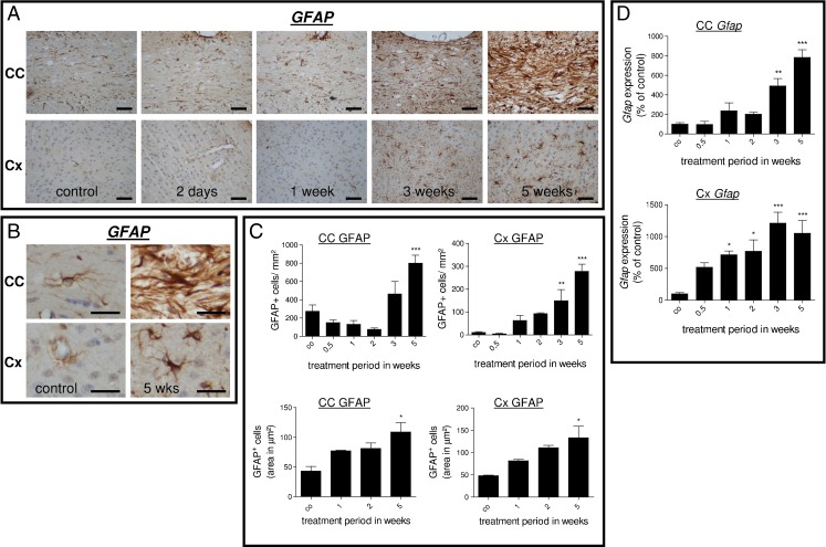
Fig. 3.
a Upper row Anti-GFAP-stained sections of the midline of the corpus callosum in control and 2 days and 1, 3, and 5 weeks cuprizone-treated animals. Note the severe astrocytosis at week 5. Lower row Anti-GFAP-stained sections of the cortex in control and 2 days and 1, 3, and 5 weeks cuprizone-treated animals. Note the moderate increase in GFAP+ cell numbers at week 5. b Micrographs in higher magnification of GFAP+ cells within the midline of the corpus callosum and cortex regions in control animals and after 5 weeks cuprizone treatment. Note the pronounced increase in number and size after 5 weeks within the midline of the corpus callosum. c Upper row Results of GFAP-expressing cell quantification within the corpus callosum and cortex regions. Lower row Morphometric quantification of GFAP-expressing cell area within the corpus callosum and cortex regions. Note that the extent of astrocyte hypertrophy is comparable in the affected white matter corpus callosum and the gray matter cortex region. d Results of Gfap gene expression analysis of the entire corpus callosum or cortex region. Each bar represents the averaged fold induction over untreated control mice of at least four mice per time point (±SEM). *p ≤ 0.05; **p ≤ 0.01; ***p ≤ 0.001 treatment vs. control. Scale bars, 50 μm (a) and 15 μm (b)

