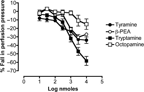
Figure 9.

Comparison of the dose–response curves for the falls in perfusion pressure of rat isolated perfused mesenteric vascular beds in response to tyramine (n= 4), β-PEA (n= 8), tryptamine in the presence of ritanserin (100 pM) (n= 7) and octopamine (n= 4). The perfusion pressure was raised by pre-constriction with phenylephrine (10 µM), which was continuously perfused for the duration of the experiment. Falls in perfusion pressure are shown as negative values, expressed as a percentage of the increase in perfusion pressure to phenylephrine. Each point represents the mean ± SEM.
