
Figure 4.
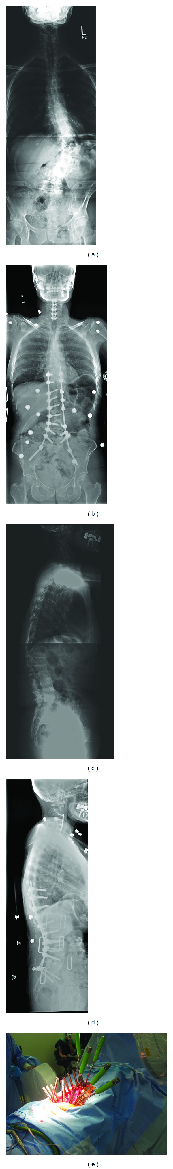
Case example showing a T9 to Iliac MIS fusion with interbody grafts at L2-S1. (a) and (b) Pre- and postoperative AP, and (c) and (d) Pre- and postoperative lateral 36” X-Ray images. (e) Intraoperative view.
Official websites use .gov
A
.gov website belongs to an official
government organization in the United States.
Secure .gov websites use HTTPS
A lock (
) or https:// means you've safely
connected to the .gov website. Share sensitive
information only on official, secure websites.
Case example showing a T9 to Iliac MIS fusion with interbody grafts at L2-S1. (a) and (b) Pre- and postoperative AP, and (c) and (d) Pre- and postoperative lateral 36” X-Ray images. (e) Intraoperative view.