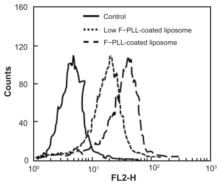
Figure 3.
Cellular association of F–PLL or low F–PLL-coated liposomes labeled with DiI in KB cells analyzed by flow cytometry after a 1-hour incubation.
Notes: The control indicates the autofluorescence of untreated cells. Each analysis was generated by counting 104 cells.
Abbreviations: DiI, 1,1′-dioctadecyl-3,3,3′,3′-tetramethylindocarbocyanine perchlorate; F-PLL, folate-poly(L-lysine).

