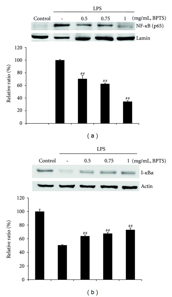
Figure 4.

Effects of BPTS on LPS-induced activation of NF-κB (p65) and degradation of I-κBα. Cells (5 × 10⁵ cells/mL) were treated with BPTS (0.5–1 mg/mL) for 1 h, followed by continuous incubation with LPS (1 μg/mL) for 15 min. Control cells were incubated with vehicle alone. Nuclear extracts for NF-κB were prepared as described in the methods section. Western blot analysis was performed for determination of protein levels of NF-κB (p65 subunit) (a) and I-κBα (b). The blots shown are representative of three blots yielding similar results. NF-κB (p65) versus Lamin A/C (a) and I-κBα versus β-actin (b) were measured via densitometry. Data represent the mean ± S.D. from three separate experiments. ## P < 0.01 and ### P < 0.001 indicate significant differences from the LPS-induced group.
