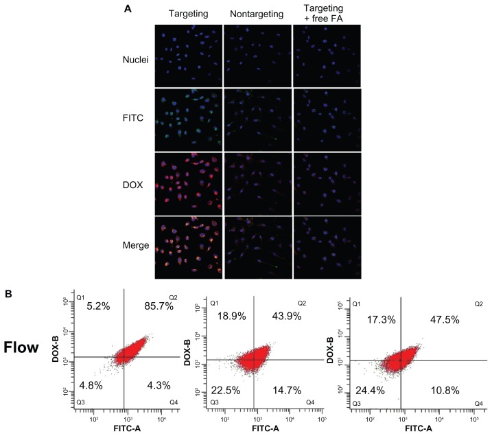
Figure 7.
Four upper panels: laser confocal microscopic images of cells incubated with D-FPPP/FITC (targeting), D-NPPP/FITC (nontargeting), and D-FPPP/FITC in the presence of FA (1 mg/L) (targeting + FA).
Notes: Bottom panel: flow cytometric measurement of DOX and/or FITC positive cells. Nuclei: stained blue with Hoechest 33342 (Aldrich); Red florescence: DOX; Green fluorescence: SCR-FITC. Incubation time: 4 hours. Dose: 50 nM siRNA. N/P = 30. DOX loading contents: 4.56% in D-FPPP and 5.14% in D-NPPP. Q1: DOX florescent cells; Q2: DOX and FITC florescent cells; Q3: nonflorescent cells; Q4: FITC florescent cells.
Abbreviations: DOX, doxorubicin; FA, folate; FITC, fluorescein isothiocyanate; D-FPPP/FITC, DOX and FITC labeled siRNA loaded folate–poly(ethylene glycol)– poly(ethylene imine)–poly(ɛ-caprolactone) micelle; D-NPPP/FITC, DOX and FITC labeled siRNA loaded nontargeted poly(ethylene glycol)–poly(ethylene imine)– poly(ɛ-caprolactone) micelle.

