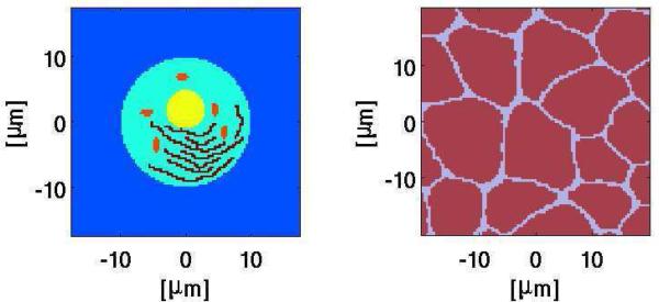
Fig. 1.
System model geometries. Left: Single cell with organelles; described elsewhere [9]. The long, inter-connected ”wiggly” black structures represent the endoplasmic reticulum (ER); the nucleus is the off-center yellow circular structure, and the five small elongated dark red structures are mitochondria. The nuclear envelope and mitochondrial membranes are double while the plasma and ER membrane are single, all with appropriate resting potential sources. Right: In vivo, multicellular with irregular shapes and sizes but no organelles; described elsewhere. [10], [11]. Papers with modeling details are all now accessible publicly [9]–[11].

