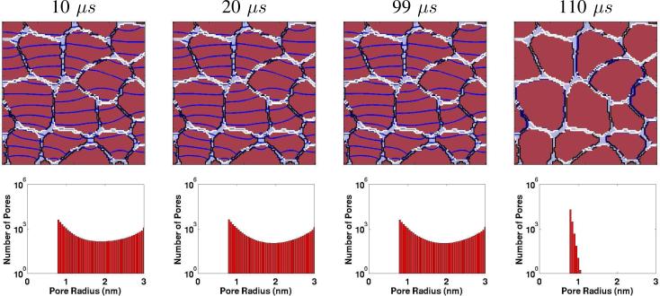
Fig. 5.
Multicellular (in vivo) model response to the IRE pulse [7]. Upper row of four panels: Spatial distributions of EP sites (WHITE regions; >1013 pores/m2) and equipotentials (DARK BLUE lines/curves). EP is mostly confined to membrane regions perpendicular to the applied field pulse (parallel to the equipotentials). Throughout the equipotentials are not parallel, due to the spatial variation in PM conductance and spatially heterogeneous EP sites. Note that the right-most panel has no equipotentials, consistent with the zero applied field at t = 110 μs. Lower row of four top/bottom panel pairs: Here, the PM pores are significantly larger (some 3 nm), but are fewer in number compared to the nsPEF case. Also note that the right-most panel shows a significantly contracted pore population. The pores have not disappeared 10 μs after the pulse ceases, but have shrunk in size to yield a thermalized (broadened) size distribution with a peak at 0.8 nm (cf. Fig. 4 at 15 ns).

