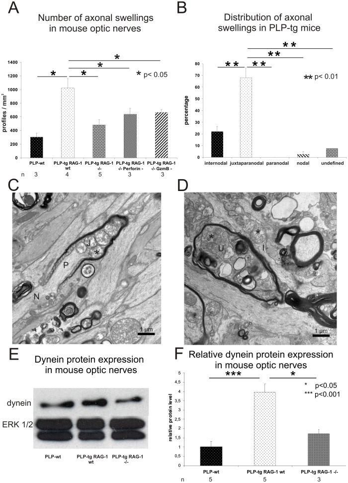
Figure 2. The occurrence of axonal swellings depend on lymphocytes, cytotoxic perforin or granzyme B and are located at juxtaparanodal aspects.
A) Number of axonal swellings (Bielschowsky’s silver impregnation, optic nerve) in PLP-wt (n = 3), PLP-tg RAG-1 wt (n = 4), PLP-tg RAG-1−/− (n = 5) mice and in PLP-tg mice devoid of cytotoxic perforin (n = 3) or granzyme B (n = 3). RAG-1 deficiency and lack of cytotoxic perforin or granzyme B in PLP-tg mice leads to a significant reduction of axonal swellings (p<0.05, one-sided student’s t-test). B) Quantification of axonal swellings in 3 PLP-tg RAG-1 wt mice by electron microscopy displays that the majority of these abnormalities are located at the juxtaparanodal region while only few abnormally organized structures are located at other domains of the myelinated fiber. Note that the paranode is not abnormally organized. C, D) Electron microscopy of PLP-tg mouse optic nerves identifies abnormal juxtaparanodal and internodal profiles, containing mitochondria (asterisks), often with an appearance reminiscent of degeneration. An axonal swelling in an undefined region is marked by an “U”. N: Node of Ranvier; P: Paranode; J: Juxtaparanode; I: Internode. E, F) Western blot analysis of optic nerves of PLP-wt, PLP-tg RAG-1 wt and PLP-tg RAG-1−/− mice. E) In this example with one individual for each genotype, dynein levels are elevated in PLP-tg RAG-1 wt mice compared to PLP-wt. RAG-1 deficiency leads to dynein levels in PLP-tg mice comparable to those found in PLP-wt mice. ERK1/2 was used as loading control. F) Quantitative Western blot analysis using densitometry and 3–5 individuals per genotype confirm the elevated dynein levels in PLP-tg RAG-1 wt mice compared to PLP-wt. RAG-1 deficiency leads to dynein levels in PLP-tg mice comparable to those found in PLP-wt mice.

