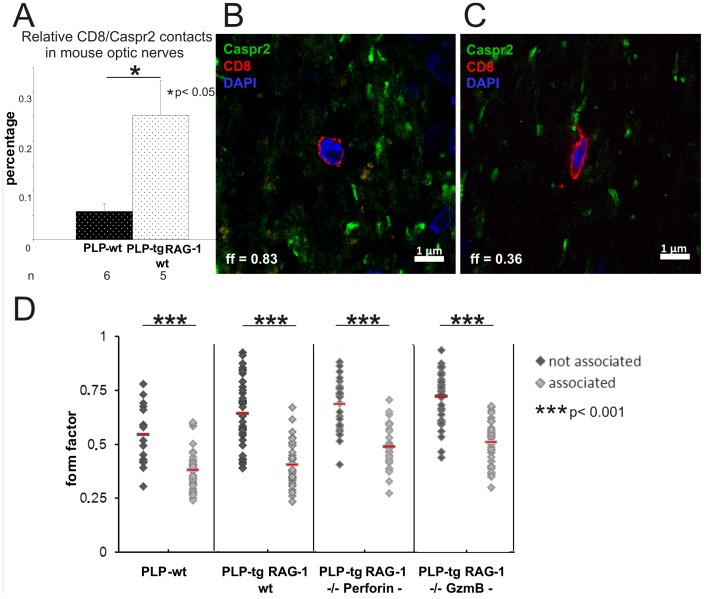
Figure 3. CD8+ T-lymphocytes contact juxtaparanodes where they acquire a spindle-shaped form.
A) Relative number of CD8/Caspr2 contacts in PLP-wt (n = 6) and PLP-tg (n = 5) mouse optic nerves. In PLP-tg mice, significantly more CD8+ T-lymphocyte-to-juxtaparanode contacts are visible than in the PLP-wt mice. B, C) Double immunofluorescence in mouse optic nerve for CD8+ T-lymphocytes (red) and Caspr2+ juxtaparanodal regions (green). Cell nuclei are stained blue with DAPI. T-lymphocytes devoid of cell contact to Caspr2+ structures are preferentially rounded (B) while T-lymphocytes attached to Caspr2+ structures are spindle shaped (C). D) Dot-plot showing form factor values of individual T-lymphocytes in PLP-wt, PLP-tg and PLP-tg mice without perforin or granzyme B. At least 16 Caspr2-associated and not-associated T-lymphocytes were investigated in each genotype (n = 2–3). Independent of the genotype, CD8+ T-lymphocytes display a low form factor representing a spindle-like shape when associated with the juxtaparanodal region.

