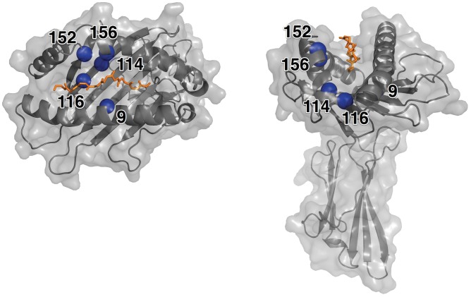
Figure 1. Structure of the HLA*A2∶01 Molecule.
The structure of the HLA-A*02∶01 (PDB id = 1AKJ) molecule shown with bound peptide backbone (chain C, orange). The positions of the five high-risk residues on the HLA molecule are represented by a blue sphere and labeled. The molecular surface of the molecule is shown in gray.

