
Figure 4. Detection of tyrosine-nitrated proteins in iNOS-overexpressed transgenic mouse retina.
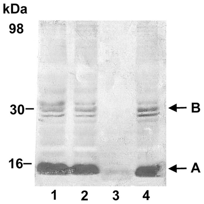
The total proteins were electrophoresed on 15% polyacrylamide gel and were probed with rabbit polyclonal anti-nitrotyrosine and biotinylated goat anti-rabbit IgG antibodies. Following enhancement with ABC kit, chromogenic visualization was used for the detection. Enhanced chemiluminescence (ECL)-based visualization was also carried out routinely for the comparison. Western blot analyses were carried out in triplicate and representative results are shown. Lane 1: heterozygote line a; lane 2: heterozygote line b; lane 3: C57BL/6 control; and lane 4: homozygote. Although there are several low-intensity tyrosine-nitrated bands in the background, the major nitrated protein pattern appears to be similar in all zygotes, with a major band near 16 kDa (A: nitrated cytochrome c monomer) and a doublet near 30 kDa (B: nitrated cytochrome c dimer). Further confirmation of these bands is presented in Figure 6.