
Figure 6. Immunohistochemical localization of tyrosine-nitrated proteins in transgenic mouse retina.
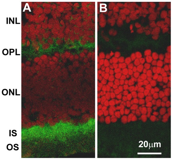
Antibodies used for the confocal immunolocalization are rabbit polyclonal anti-nitrotyrosine and Alexa Fluor 488-conjugated goat anti-rabbit IgG (green). Propidium iodide (red) was used for the nuclear staining. The intense localization was seen specifically and uniformly in the photoreceptor inner segments (IS) and outer plexiform layer (OPL). These locations are known mitochondria-rich areas. A: retina section from homozygote; B: retina section from control. Retina layers labeled are: INL: inner nuclear layer; OPL: outer plexiform layer; ONL: outer nuclear layer; IS: photoreceptor inner segments and OS: photoreceptor outer segments.