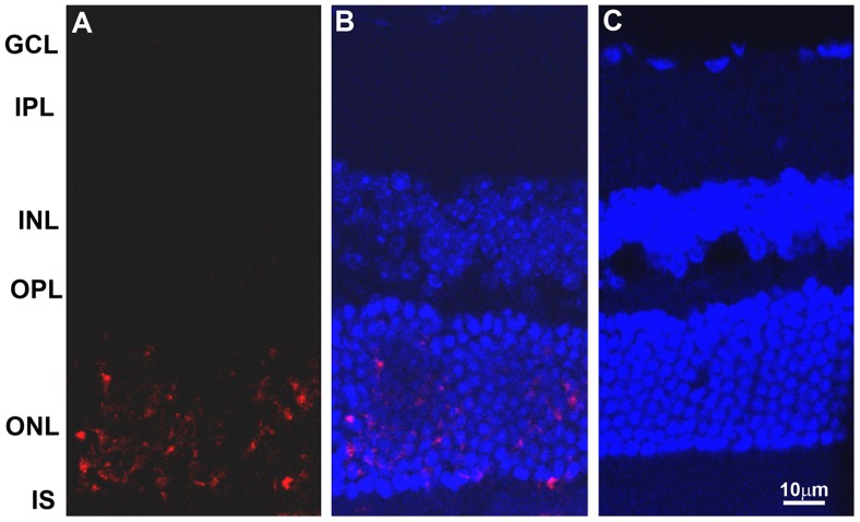
Figure 7. Detection of apoptotic cells in retina of iNOS transgenic mice.
Retinal apoptosis was detected using In Situ Cell Death Detection Kit, TMR (trimethyl rhodamine) red, and the free 3′-OH from the DNA strand breaks are detected by modified nucleotides in an enzymatic reaction (TUNEL reaction). A: Apoptotic cells without nuclear staining; B: Apoptotic cells after DAPI staining; and C: C57BL/6 control retinal section with both TUNEL and DAPI staining. A large number of apoptotic cells were found specifically in the outer nuclear layer and none in the other retinal layers. A small number of apoptotic cells (3–4 cells) were routinely seen in the entire segment of control retinal sections. Substantially more apoptotic cells are visible in A without DAPI staining, since following merging with DAPI, some apoptotic cells were buried under the ONL nuclear staining.

