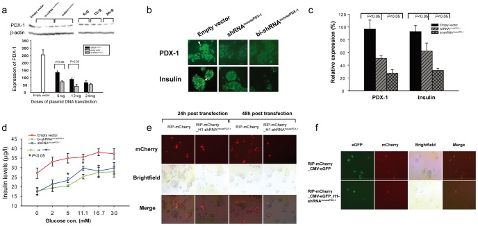
Figure 4. bi-shRNAmousePDX-1 knockdown of mPDX-1 expression in β TC-6 cells in vitro inhibits insulin expression and secretion.
Comparison of efficacy of bi-shRNAmousePDX-1, shRNAmousePDX-1 or empty vector in inhibition of PDX-1 expression in β TC-6 cells is shown using western blot (a) and cell immunostaining (top panel, b as indicated by arrow). Insulin expression and glucose stimulated secretion in response to knockdown of PDX-1 is shown by cell immunostaining (bottom panel, b as indicated by arrow) (×200) and ELISA assay (d), respectively. Each experiment was repeated five times. PDX-1 and insulin expression in immunostained β TC-6 cells are quantified (c). PDX-1 expression affected the RIP-directed reporter expression (RIP-mCherry) in βTC-6 cells (e and f). The cells expressing mCherry (red) and GFP (green) were visualized and photographed using fluorescence microscopy (e and f). (×200).

