Figure 1.
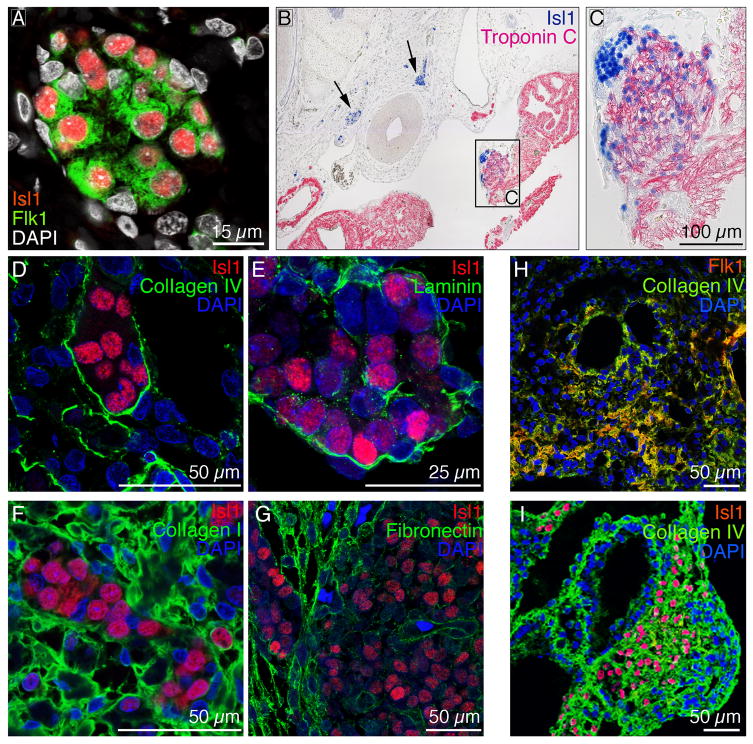
(A) Immunofluorescence staining of 9 week human hearts reveals that endogenous Isl1+ CPCs (red) also express Flk1 (green). DAPI-stained cell nuclei are shown in white. (B-C) Immunohistochemistry shows that endogenous Isl1+ CPCs are localized in clusters (B, see arrows). Differentiating CPCs down-regulate Isl1 (blue) and up-regulate mature cardiac markers such as troponin C (pink) (C). (D-G) ColIV (D; green) and laminin (E; green) are tightly expressed around the CPC clusters, whereas ColI (F; green) and fibronectin (G; green) are predominantly expressed outside the niche within the myocardium. Cell nuclei are shown in blue (DAPI). (H-I) Flk1+ and Isl1+ CPCs (red) co-localized with ColIV (green) in differentiating mouse embryoid bodies. DAPI-stained nuclei are shown in blue.

