Figure 2.
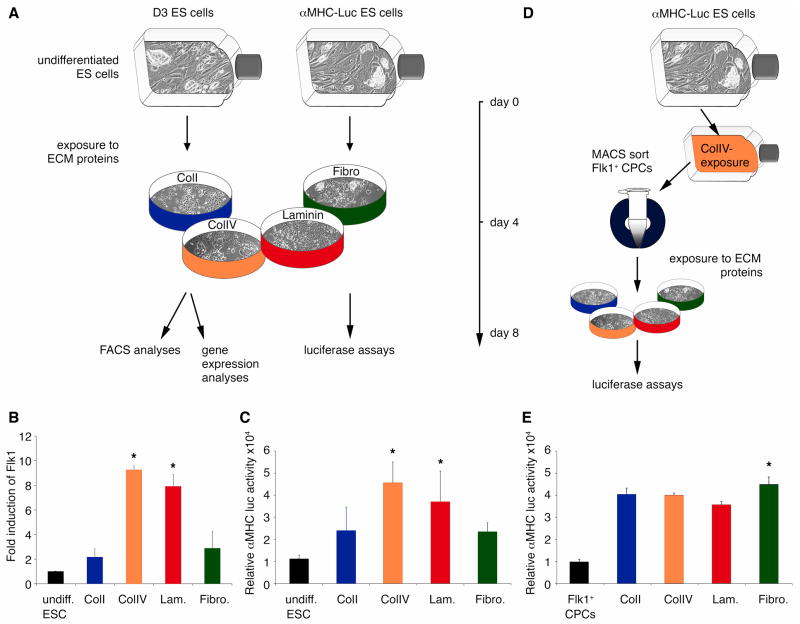
(A) D3 ES and alpha-MHC-Luc ES cells were cultured for 4 days on ColI (blue), ColIV (orange), laminin (Lam.; red) or fibronectin (Fibro.; green). (B) Quantitative real-time PCR demonstrates a significant increase in Flk1 expression in differentiating ES cells cultured on ColIV or laminin *P<0.05 versus undifferentiated (undiff.) ES cells; ColI- and Fibro-exposed cultures. (C) Luciferase assays show significantly higher alpha-MHC-luciferase activities in ColIV- and Lam.-exposed cultures. *P<0.05 versus undiff. ES cells; ColI- and Fibro-exposed cultures. (D) alpha-MHC-Luc ES cells were cultured for 4 days on ColIV before Flk1+ CPCs were MACS-sorted. Flk1+ CPCs were then exposed for 4 days to ColI (blue) or ColIV (orange), Lam (red) or Fibro (green). (E) Luciferase assays show significantly higher alpha-MHC-dependent luciferase activity in Fibro-exposed cultures. *P<0.05 versus alpha-MHC-Luc ES cell-derived Flk1+ CPCs; ColI-, ColIV- and Fibro.-exposed cultures.

