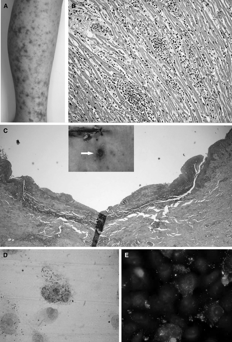
Figure 1.
(A) Maculopapular rash with areas of central necrosis violacea. (B) Lymphocytic myocarditis with vasculitis in small vessels. (C) Skin section of the lesion (Tache noire), showing necrosis and leukocytoclastic. (D) Gimenez staining of VERO E 6 cells infected with isolated Rickettsia. (E) Immunofluorescense of VERO E6 cells infected with isolated R. rickettsii.

