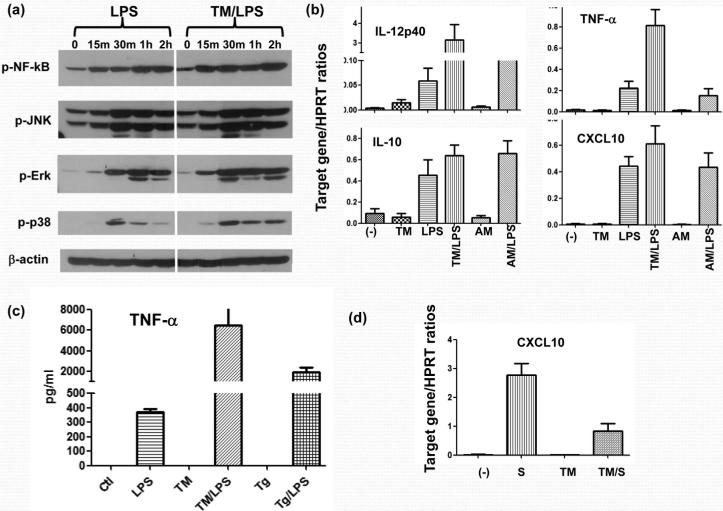
Figure 1.
ER stress enhances TLR4-induced macrophage pro-inflammatory immune response. RAW cells were first incubated with Tunicamycin (TM) or Antimycin (AM), followed by LPS stimulation, as described in Material and Methods. Cells were harvested after 15 min, 30 min, 1 h or 2 h after LPS stimulation, and cell lysates were analyzed for NF-kB, JNK, Erk and p38 phosphorylation, as well as β-actin protein levels by Western blots (a). In a separate experiment, RAW cells were harvested after 4 h-long LPS stimulation and total RNA was isolated and subjected to qRT-PCR analysis (b). Bone-marrow-derived macrophages were stimulated first with ER stress agent Tm or Tg, followed by 24h LPS stimulation. Culture supernatants were harvested and TNF-a levels were measured by ELISA (c). Mouse hepatoma cell line Hepa-1, was stimulated with either control (-) or LPS-stimulated (S) RAW cell culture supernatant w/ or w/o pre-incubation with Thapsigargin (Tg). Cells were harvested after 4 h of stimulation and total RNA was isolated and subjected to qRT-PCR analysis (d). Representative results of 3 separate experiments.

