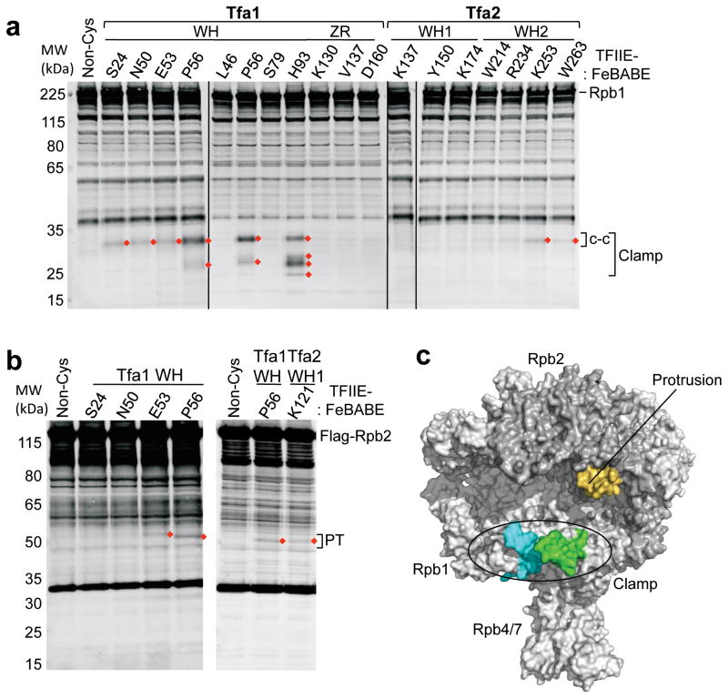
Figure 2.
Mapping of TFIIE-FeBABE cleavage in Pol II. (a) Hydroxyl radical-mediated cleavage in Pol II subunit Rpb1 by the indicated TFIIE-FeBABE variants. PICs were formed at the HIS4 promoter using yeast nuclear extracts that were supplemented with recombinant TFIIE-FeBABE. Rpb1 cleavage products were visualized by Western blot using an antibody against the N-terminus of Rpb1 6. Red diamonds indicate reproducibly observed cleavage products compared to a reaction with a non cysteine-containing TFIIE (Non-Cys; lane 1). Brackets on the right indicate cleavage in the clamp and the coiled-coil domain (c-c). Bands in the Non-Cys control (lane 1) correspond to full length Rpb1 and non-specific reactivity of the primary/secondary antibodies with other proteins in the reaction. (b) Rpb2 cleavage using the indicated TFIIE-FeBABE derivatives. The right bracket indicates cleavage in the protrusion domain (PT). (c) Summary of the TFIIE-FeBABE mediated cleavages on Pol II. All cleavage events that are summarized in Table S1 were mapped on the Pol II surface and 10 residues on either side of the calculated cleavage sites are colored (green = cleavage in the Rpb1 clamp coiled-coil domain generated by FeBABE linked to residues in the TFIIE dimerization domain; cyan = cleavage in the Rpb1 clamp head by FeBABE conjugated to residue Tfa1 His93; orange = cleavage in the Rpb2 protrusion domain by FeBABE at Tfa1 Glu53, Pro56, and Tfa2 Lys121).

