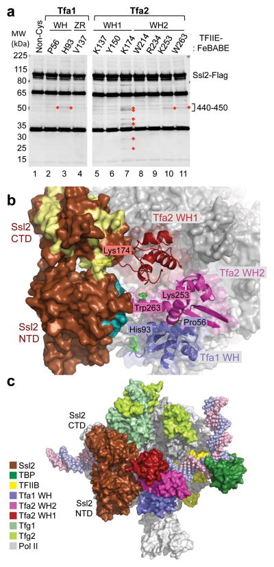
Figure 6.
Orientation of the TFIIH subunit Ssl2/XPB in the PIC. (a) Cleavage in Ssl2 by TFIIE-FeBABE variants. PICs were formed with nuclear extracts containing Ssl2-Flag and supplemented with TFIIE-FeBABE. Cleavage was induced by addition of H2O2 and products were visualized by Western blot using an anti-Flag antibody. Reproducibly observed cleavage products are highlighted with red diamonds. The majority of TFIIE-FeBABE variants that cut Ssl2 cleaved in a region spanning residues 440–450 on the Ssl2 N-terminal domain. (b) PIC model containing TFIIE and Ssl2. Ssl2 was positioned in the PIC based on hydroxyl-radical cleavage by FeBABE linked to various positions in TFIIE (shown as green, or yellow for Lys174). Ssl2 cleavage by residues in the TFIIE dimerization domain is colored teal, cleavage by Tfa2 residue Lys174 is colored pale yellow. (c) Model of the PIC containing Ssl2. Different components of the PIC are colored according to the color-legend.

