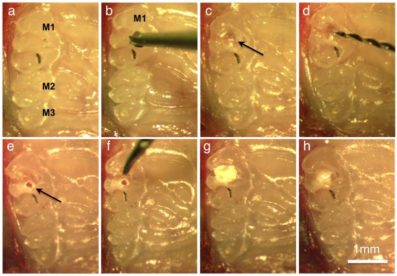
Figure 1.
Procedure of pulp exposure, capping, and restoration. (a) Occlusal view of the first (M1), second (M2) and third (M3) molars. In all images the palatal side of the arch is on the right. (b) Position of the carbide bur on the centre of the first molar (M1). (c) Small cavity (arrow) on centre of the occlusal surface. (d) Endodontic hand file used to mechanically expose the pulp. (e) Pulp exposure (arrow). (f) Probe used to apply the MTA. (g) Pulp capping with MTA. (h) Cavity restoration with resin composite.

