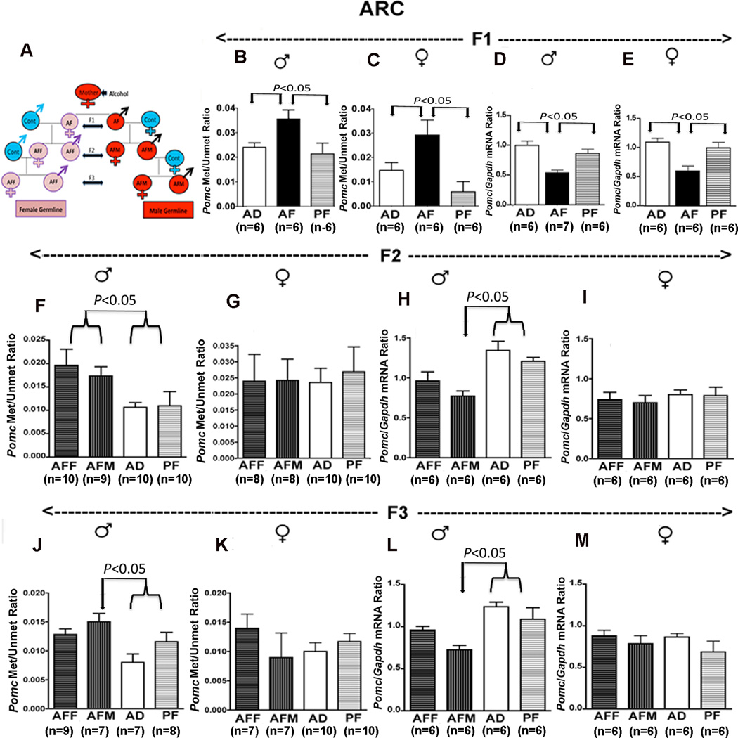
Figure 5. Transgenerational changes in hypothalamic Pomc gene methylation, expression and functions after alcohol feeding in pregnant female rats (F0).
A schematic diagram to indicate how F1, F2 and F3 male germline (AFM) and female germline (AFF) fetal alcohol-exposed offspring were generated (A). Methylation-to-unmethylation ratio of the CpG pairs in the −81 to −154 region of the Pomc promoter in ARC tissues of F1 (B, C), F2 (F, G) and F3 (J, K) male and female rat offspring of male and female germlines. Pomc mRNA levels in the ARC tissues of F1 (D, E), F2 (H, I) and F3 (L, M) male and female rat offspring from different germlines. Data presented are mean ± s.e.m. The number of animals was indicated within brackets under each histogram. Pomc methylation and unmethylation ratio data were analyzed using using Kruskal-Wallis ANOVA followed by Dunn's posttest. Pomc mRNA levels data were analyzed using a one-way ANOVA followed by the Student-Newman-Keuls posthoc test (see Table S6 in the Supplement). The significance of difference between groups was identified by a bar above histograms.

