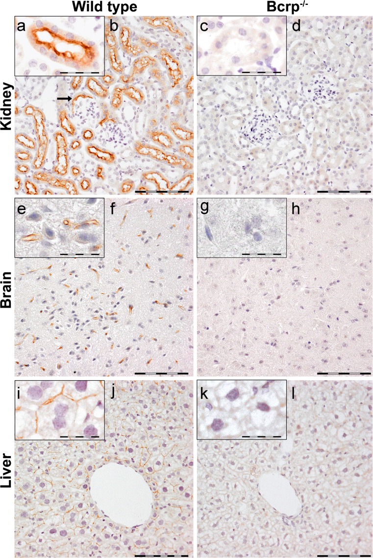
Fig. 2.
Representative immunohistochemical images of Bcrp distribution in wild-type and Bcrp−/− (negative control) mouse kidney (a–d), brain tissue (e–h) and liver (i–l). Because of an absence of gender difference in localization, only organs from female mice are shown. Perfusion-fixed, paraffin embedded organ sections were incubated with a primary antibody against mouse Bcrp (BXP-9). DAB chromogene staining (brown) visualizes Bcrp localization. The sections were counterstained with Mayer’s hematoxylin. Bars 100 μm in (b, d, f, h, j, l). a, e, i represent details of Bcrp positivity and corresponding negative controls (c, g, k); bars in these insets 50 μm. Bcrp was present in the brush border of proximal tubule and in the brush border of Bowman’s capsules (arrow) of the kidney and in the capillaries of the brain and liver

