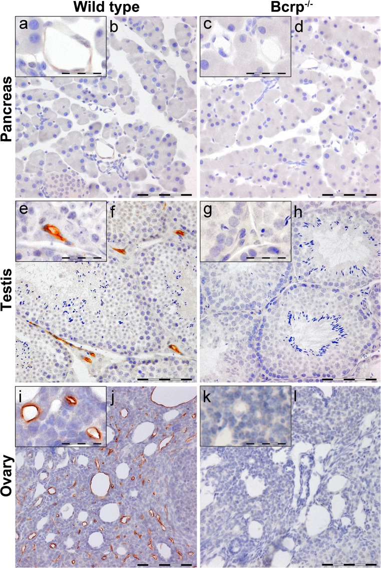
Fig. 4.
Representative immunohistochemical images of Bcrp distribution in wild-type and Bcrp−/− (negative control) mouse pancreas (female, a–d), testis (e–h) and ovary (i–l). Perfusion-fixed, paraffin embedded organ-sections were incubated with a primary antibody against mouse Bcrp (BXP-9). DAB chromogene staining (brown) visualizes Bcrp localization. The sections were counterstained with Mayer’s hematoxylin. Bars 100 μm (b, d, f, h, j, l). a, e, i represent details of Bcrp positivity and corresponding negative controls (c, g, k); bars in these insets 50 μm. Blood vessels in the testis and ovary showed distinct Bcrp positivity, whereas in the pancreas, they showed very subtle Bcrp staining

