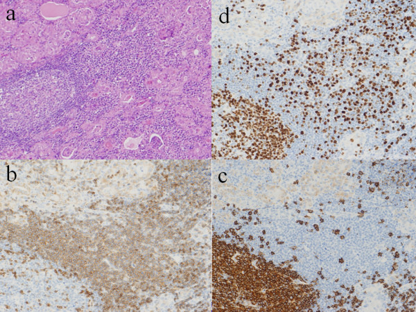
Figure 3.

Microscopic findings and immunohistochemical staining. (a) Low-power view of histological examinations revealed massive infiltration of small monotonous lymphocytes, which were difficult to distinguish tumor cells from reactive lymphocytes in Hashimoto’s thyroiditis. (Hematoxylin and eosin staining, X100). (b) Immunohistochemical staining showed that tumor cells had T-cell markers for CD3, (X400). (c) Immunohistochemical staining by CD20 showed infiltrated lymphoid cells had B-cell markers, (X400). (d) MIB staining. MIB 1 index was as high as 60% in high-power fields, (X400).
