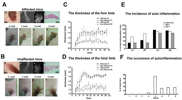
Figure 1.
Phenotype of paw morphology in unaffected and affected mice. Mouse detected by X-ray (A,b), MRI (A,c), and H&E staining (A,d). (A) (a) Clinical symptoms of edematous inflammation and necrosis of the digits (arrows). (b) Radiography of a paw in an affected mouse shows bone destruction, and the shadow shows enlarged soft tissue. (c) Affected mice exhibited indistinct signals of swollen soft tissue revealed by MRI. (d) Inflamed phalanges of H&E stains in affected mice. The arrows show hyperplasia of the epithelium. (A,e-h) Paw development in unaffected offspring from week 6 to week 9. (B,a-h) Comparison of respective phenotypes in unaffected mice. (C) Thickness of the digits in the inflamed forelimb in mice with autoinflammation and unaffected mice. (D) Thickness of the digits in the inflamed hindlimb in affected and unaffected mice. (E) Percentage of penetrance in each limb in affected mice. (F) Age of onset of clinical symptoms in affected mice. Abbreviations: LF, left forelimb; RF, right forelimb; LH, left hindlimb; RH, right hindlimb.

