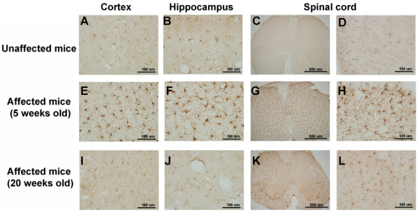
Figure 6.

Morphological microglial changes in unaffected and affected mice. Microglia were stained with an Iba-1 antibody. (Upper panel) Microglia stainings in the cortex (primary somatosensory cortex), hippocampus (dentate gyrus), and spinal cord of unaffected mice. (Middle and lower panels) Microglia stainings in respective brain areas of 5-week-old and 20-week-old affected mice.
