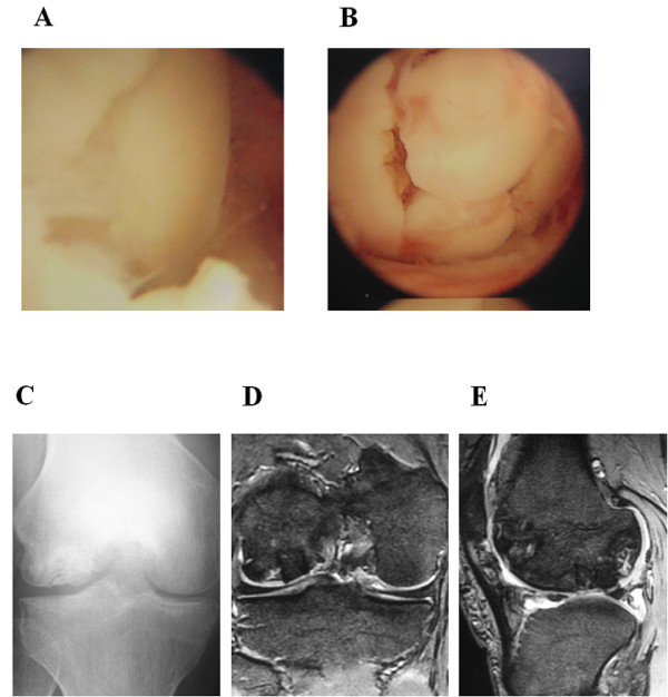
Figure 2.

Operative and postoperative findings in case 1. (A) Arthroscopy showing the lesion of the osteonecrosis at the medial condyle of the femur demonstrating the anterior margin of the femoral condylar defect. (B) Osteochondral plugs are grafted into the recipient site. (C) Anteroposterior radiograph taken one year after surgery shows less irregularity of the subchondral bone of the lateral femoral condyle and a larger sclerotic area. (D, E) Coronal and saggital MRIs taken one year after surgery also show less irregularity of the subchondral bone and the reduction of the osteonecrotic area as compared with the preoperative MR images (Figure 1B,C), although a high signal area remains slightly in the lateral and posterior aspect of the lesion.
