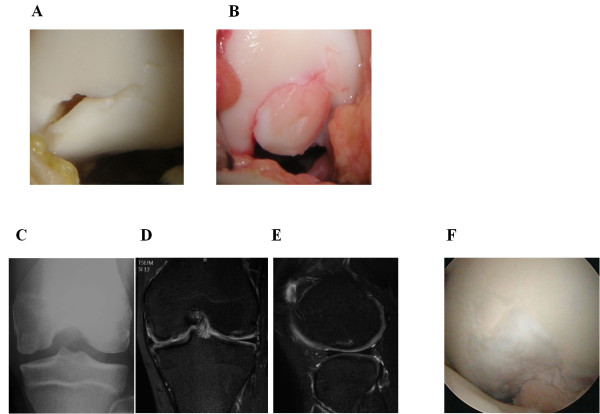
Figure 4.
Operative and postoperative findings in case 2. (A) Arthroscopy shows that the free margin of a chondral fragment still partially attached to the femoral condyle posteriorly. The anterior margin of the femoral condylar defect is seen. (B) An osteochondral plug is grafted into the recipient site. (C) Anteroposterior radiograph taken two year after surgery shows less irregularity of the subchondral bone of the medial femoral condyle. (D, E) MRI taken two years after surgery shows the restoration of the articular cartilage surface and good engraftment of the graft. (F) Second look arthroscopic finding of the transplanted site demonstrates that the lesion is covered with cartilageous tissue.

