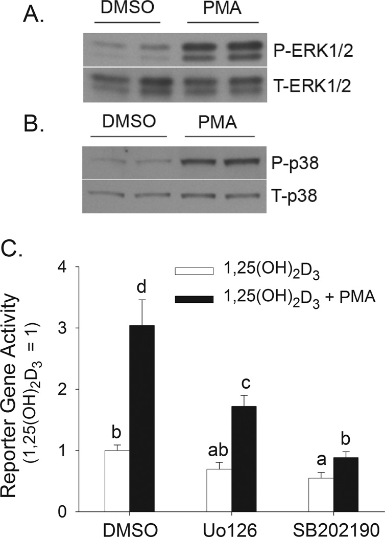
Figure 3. Contribution of PMA-induced activation of ERK1/2 and p38 kinase to 1,25(OH)2D3-induced hCYP24A1 promoter activity in differentiated Caco-2 cells.
(A,B) Differentiated Caco-2 cells were treated with vehicle (DMSO) or 100 nM PMA for 5 min. Cells were collected immediately (for ERK1/2 analysis) or 1 h after 5 min PMA treatment (for p38 kinase analysis). (A) Total (T-ERK1/2) and phospho-ERK1/2 (P-ERK1/2) levels. (B) Total (T-p38) and phospho-p38 kinase (P-p38) levels. Data are representative of three independent experiments. Two samples were analyzed per treatment. (C) Differentiated Caco-2 cells were transfected with a −298 to +74 bp human CYP24A1 promoter-luciferase construct. After transfection, cells were preincubated with inhibitors of MEK 1/2 (10 µM U0126) or p38 kinase (8 µM SB202190) for 30 minutes in the presence or absence of 100 nM PMA for the last 10 min. Inhibitor co-treatment continued with 10 nM 1,25(OH)2D3 or ethanol vehicle for an additional 4h in the absence of PMA. Data were expressed relative to the 1,25(OH)2D3-induced expression of the reporter gene (calculated as 1,25(OH)2D3/vehicle = 100) (mean±sem, n=4). The values with different letter superscripts are significantly different from one another (p < 0.05, Fisher’s protected LSD). Data are representative of three independent experiments.

