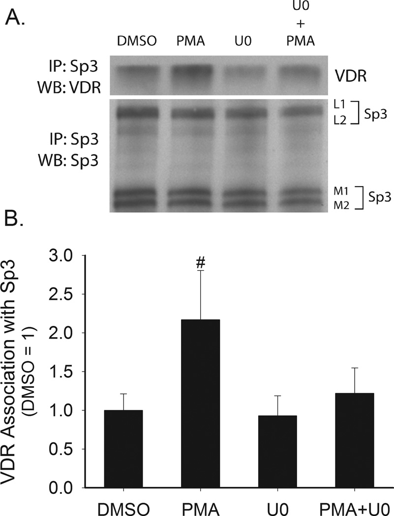
Figure 7. Effect of PMA on the interaction between Sp3 and VDR in Caco-2 cells.
Proliferating Caco-2 cells were pretreated with U0126 (10 µM,, 30 min) followed by treatment with 100 nM PMA for 5 min. Whole cell extracts were collected and Sp3 was immunoprecipitated. Immunoprecipitated proteins were analyzed by Western blot analysis for Sp3 and VDR (A) Representative Western blot. (B) Quantitative data from four repeat experiments. Data are expressed as mean±sem (n=4) of the fold change above non-treated levels. # represents significant different from all other groups (p < 0.05).

