
Figure 7.
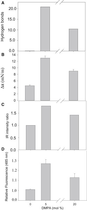
A comparison of data concerning Aβ42 binding to bilayers composed of SM/Chol (1:1 mol ratio), to which increasing amounts of anionic phospholipid (phosphatidic acid) had been added. (A) MD calculations of average H-bonds established between peptide and membrane for the last 20 ns. Data taken from Fig. 2B. (B) Langmuir balance. Increase in surface pressure of lipid monolayers (16 mN/m) of the compositions given previously due to the presence of Aβ42. Data are taken from Fig. 4B. (C) Relative change in intensity of the IR band centered at 1622 cm−1, assigned to antiparallel β-sheet vibration. Data taken from Fig. 6. (D) Changes in ThT fluorescence after 6 h incubation with Aβ42 and LUVs. Data taken from Fig. 5.