
Figure 2.
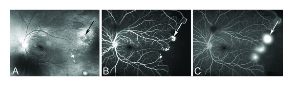
(A) Fundus photograph of new vascular formations characteristic of sickle cell disease called sea fan formations (arrow), which tend to occur in the temporal periphery. (B) Fluorescein angiogram in the arteriovenous phase shows peripheral nonperfusion and sea fan neovascularization at the border of vascularized and nonvascularized retina (arrow indicates the largest sea fan). (C) Late phase of the angiogram shows fluorescein leakage from sea fans as the dye study progresses.