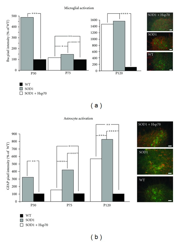
Figure 5.

rhHsp70 injections attenuate early glial cell activation. (a) The fluorescent pixel intensity of Iba1 staining for microglia was increased in the SOD1 versus WT animals at P30 and P75. Treatment with rhHsp70 appeared to reduce the expression of Iba1 at P75, but the levels were greater than WT animals. Pixel intensity was measured using Image J software. Each fluorescent pixel in the section was measured and the values were compiled and averaged across each treatment group. Histograms represent the average number of pixels in a given range of arbitrary intensity units presented as % WT. Statistical significance was determined using a two-way ANOVA at P30 and a repeated measure (mixed model) ANOVA at P75 on all of the measured values above baseline (***P ≤ 0.001; **P ≤ 0.01; *P ≤ 0.05). N = 4 for each group. Images of the lateral motor column of P75 mice that were treated with Hsp70, untreated, or WT are shown on the right. 30 μm spinal cord sections were stained with Iba-1 (red) and ChAT (green). Scale bars = 20 μm. (b) GFAP expression was the greatest in SOD1 animals as compared to WT at P30 and P75. Treatment with rhHsp70 substantially reduced the expression of GFAP in SOD1 animals. Quantification of fluorescence and statistical analysis was performed as described above. Images of the ventral horn of P75 mice that were treated with Hsp70, untreated, or WT are shown on the right. 30 μm spinal cord sections were stained with GFAP (red) and ChAT (green). Scale bars = 20 μm.
