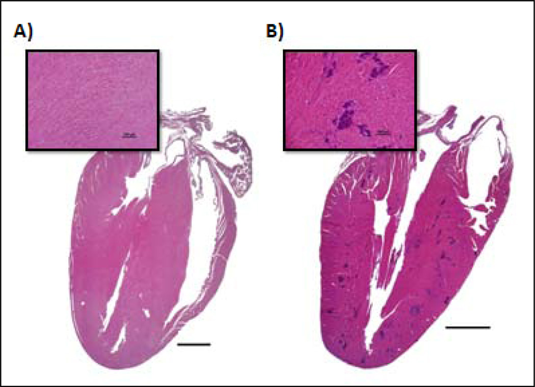
Fig. 5.
Photomicrographs comparing an untreated mouse heart to a heart with dystrophic cardiac calcification (DCC). A, Normal heart section with inset of the myocardium demonstrating no evidence of myocardial mineralization. B, Section and inset from a mouse demonstrating moderate, multifocal myocardial mineralization (intensely basophilic foci) throughout the ventricular free wall and interventricular septum characteristic of DCC. Hematoxylin and eosin staining of paraffin-embedded formalin-fixed tissue.

