
Figure 3.
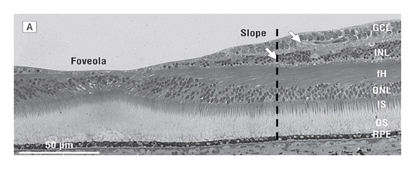
This shows histology of the human retina (after Provis and Hendrickson). Interrupted lines represent perifoveolar radial distances of 0.9 and 1.25 mm where ganglion cells begin to dominate inner retinal thickness. Maximal IA occurs at distances less than 0.9 mm [22].