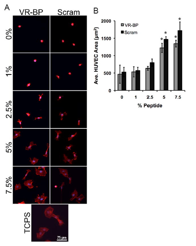
Figure 5.

Assaying for non-specific protein adsorption and surface fouling. HUVECs were seeded on SAMs presenting varied densities of VR-BP, scrambled peptide, or tissue culture polystyrene and (A) stained for their f-actin cytoskeleton (f-actin:red, nuclei:blue). (B) HUVEC projected cell area was used as a measurement of surface fouling. (Error bars represent standard error of the mean, asterisk indicates significant increase in projected cell area compared to 0% peptide, p < 0.05) % GRGDSP % GRGDSP
